


A client brought in her eight-year-old Labrador last spring for a routine senior exam. The dog’s vision was still sharp — tracking a treat across the room without hesitation — but the owner had noticed that her previous Lab had started bumping into furniture around the same age. She asked me whether there was anything in the diet that might help protect her current dog’s eyesight. Lutein came up immediately.
The conversation lasted about fifteen minutes. By the end I realized I’d covered almost every question her vet hadn’t had time to answer: what lutein actually does in the eye, why it matters more for some breeds, and where the evidence sits between “well-established” and “promising but incomplete.” That conversation is basically what this article is.
The Retina’s Built-In Light Filter: What Lutein Really Is
Lutein is a xanthophyll carotenoid — a type of fat-soluble pigment that plants produce to protect their own tissues from excessive light. Animals don’t make it. Every milligram in your dog’s or cat’s bloodstream came directly from something they ate.
What makes lutein physiologically interesting isn’t that it exists in the body — dozens of carotenoids circulate in blood plasma — it’s that the body selects it. Of the hundreds of carotenoids in a typical diet, only two (lutein and zeaxanthin) are actively concentrated in the macula and retina. The retina contains more lutein per gram than almost any other tissue in the body (Bhosale & Bernstein, 2007). That selective accumulation isn’t random. It points to a specific function.
The pigment formed by this accumulation is called macular pigment. Researchers measure its density using a value called macular pigment optical density, or MPOD. Higher MPOD means the eye has more of this protective filter in place. In humans, lower MPOD is associated with higher long-term risk of macular degeneration. Whether the same relationship holds in dogs and cats isn’t yet fully characterized — but the anatomy is close enough that researchers use dogs as models in human ocular disease studies, which tells you something (Berman et al., 2013).
How Lutein Protects the Eye: Two Distinct Mechanisms
Understanding why lutein matters means separating its two main jobs. They’re related but distinct, and conflating them leads to vague claims that don’t help you make a good decision.
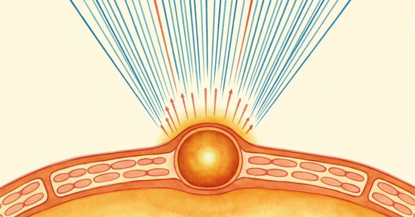
Blue-light absorption
Visible light spans a spectrum from about 380 to 700 nanometers. Blue light — the highest-energy end of the visible spectrum, roughly 430–480nm — carries the most photon energy and does the most cumulative damage to retinal tissue over time. Lutein absorbs directly in this range. It acts as a biological sunscreen for the photoreceptors behind it (Stahl & Sies, 2003).
Dogs and cats encounter meaningful blue-light exposure daily: outdoor sunlight, reflected light off water or pavement, and — increasingly — indoor LED and screen light. The retinal tissue that processes this light, particularly the cone-rich central region, is metabolically active and vulnerable. Lutein positioned in the macular pigment intercepts a portion of incoming blue-light photons before they reach the photoreceptors.
How much protection? That’s harder to quantify. Studies in healthy humans suggest a 20–30% reduction in blue-light transmittance at normal macular pigment densities (Hammond et al., 1997). Similar measurements in dogs haven’t been published at the same scale, but the optical physics are the same. The pigment filters what it filters, regardless of species.
Antioxidant activity
Photoreceptors burn through oxygen at an extraordinary rate. The high metabolic demand — combined with constant light exposure — generates reactive oxygen species, which are basically cellular exhaust that damages nearby tissue over time.
Lutein neutralizes these radicals. Its molecular structure allows it to donate electrons to unstable oxygen molecules, breaking the chain reaction before it damages photoreceptor membranes or the proteins that make vision possible. This antioxidant role is particularly relevant in the outer retina, where rod and cone outer segments turn over constantly and oxidative byproducts accumulate (Bhosale & Bernstein, 2007).
This isn’t unique to lutein — many antioxidants do similar work — but lutein is positioned exactly where oxidative stress is highest. That positioning matters more than total antioxidant capacity.
What Studies in Pets Actually Show
Here’s where I try to be honest rather than enthusiastic.
The strongest evidence for lutein’s eye benefits comes from human research. The AREDS2 study — a large randomized trial — found that lutein supplementation (10mg/day) reduced risk of advanced age-related macular degeneration by about 18% compared to beta-carotene in participants with existing intermediate AMD (AREDS2 Research Group, 2013). That’s a solid result, in a specific population, for a specific outcome.
In dogs, the picture is more limited but not empty. Bjerkås et al. (2002) demonstrated that dietary lutein increased MPOD in healthy Beagles, confirming that supplemented lutein does cross from bloodstream to retinal tissue. That’s an important step — it means lutein in food or supplements isn’t just metabolized and discarded; it actually gets where it needs to go.
Malone et al. (1995) showed that carotenoid profiles in canine plasma respond to dietary changes within weeks, suggesting the system is responsive and not static. A dog eating a diet low in lutein will have lower retinal lutein over time, and can replenish it.
For cats, direct lutein studies are sparse. Cats evolved as strict carnivores consuming prey whose own tissue contained whatever carotenoids those prey had absorbed. Commercial dry food is often extremely low in bioavailable lutein. Whether chronic low dietary lutein leads to measurable changes in feline MPOD or long-term visual outcomes — nobody has published a definitive answer on that yet.
What I tell clients: the mechanistic case for lutein is solid. The clinical trial evidence in pets is limited. That puts lutein in the “reasonable to include, honest about ceiling” category rather than “proven to prevent eye disease.”
Where Lutein Comes From in Your Pet’s Diet
In the wild, dogs eating prey and plant material would encounter lutein through muscle tissue, organ meat (especially liver), and incidental plant ingestion. Cats, as obligate carnivores, would derive lutein almost entirely from organ tissues of prey species.
Commercial pet foods are variable. Dry kibble processing is hard on heat-sensitive nutrients — the high-temperature extrusion process degrades carotenoids, including lutein (Fernández-García et al., 2012). Wet foods fare better, particularly those with whole-ingredient organ meats. Raw or gently cooked diets, depending on ingredients, may preserve more native lutein.
Common lutein-rich ingredients you might see in higher-quality pet foods:
- Egg yolks — one of the most bioavailable lutein sources; lutein from egg is better absorbed than from plant sources because of fat co-ingestion (Wenzel et al., 2010)
- Marigold flower extract (tagetes extract) — the most common supplemental source; high in both lutein and zeaxanthin
- Liver and organ meats — contain meaningful carotenoid content depending on what the source animal ate
- Dark leafy greens — significant in human nutrition but low-utility for cats, and typically low in standard pet food ingredient panels
Bioavailability from supplement sources matters too. Lutein is fat-soluble, meaning it absorbs much better when consumed alongside fat. A lutein supplement given to a cat that just ate dry kibble in a fat-sparse meal will absorb at a fraction of the rate compared to lutein consumed with a fat-containing wet food (Bhosale & Bernstein, 2007). If you’re using a supplement, timing it with a meal that contains fat makes a real difference.
Lutein in Supplements: Reading Labels Without Getting Lost
The supplement market around pet eye health is crowded. Lutein appears in formulas alongside zeaxanthin, bilberry extract, astaxanthin, and a long list of antioxidants. A few things worth paying attention to:
Check the source. Most commercial lutein is derived from marigold flowers (Tagetes erecta), which is the most studied and reliable source. Some cheaper products list “lutein” without specifying derivation — not necessarily a problem, but worth noting.
Look at the unit. Human lutein doses in research are typically 10–20mg/day. For dogs, published feeding studies used ranges from about 5mg to 20mg daily. There isn’t an established veterinary daily value, but seeing a dose under 1mg in a product probably means eye health is more of a marketing angle than a formula priority.
Fat inclusion matters. Lutein supplements in soft chews or oil-based formats tend to absorb better than those in dry tablet form, assuming there’s fat in the vehicle.
Context in combination formulas. Lutein often appears alongside lactoferrin in tear and eye support products. These ingredients address different things — lutein acts in the retina, while lactoferrin has antimicrobial and anti-inflammatory roles in the tear film and ocular surface. When both appear together, they’re typically targeting different aspects of ocular health rather than duplicating each other. Similarly, L-lysine occasionally shows up in eye-adjacent products because of its antiviral role in feline herpesvirus-related eye symptoms — again, a different mechanism from lutein’s retinal function.
For dogs prone to tear staining, the Petterm Tear Stain Powder includes lutein as part of a multi-ingredient approach to periocular health. The lutein component there targets retinal antioxidant support rather than tear staining directly — the two concerns are anatomically adjacent but mechanistically separate. If your dog has significant oxidative stress in the ocular environment, addressing the inner eye and the outer eye simultaneously makes sense as a support strategy.
Frequently Asked Questions
Does lutein help prevent cataracts in dogs? Cataracts involve clouding of the lens, not damage to the retina. Lutein concentrates in the macula and retina, not the lens. There’s no strong evidence it helps prevent or slow cataracts. The conditions are related to oxidative stress, but lutein isn’t positioned where cataracts form.
My cat is on a dry food diet. Should I worry about lutein deficiency? Most commercial dry cat foods have lower bioavailable lutein than a diet containing fresh organ meats. Whether that creates a meaningful deficiency over a cat’s lifetime isn’t established in the literature. If you’re already adding a supplement for another reason, choosing one that includes lutein or zeaxanthin is a reasonable choice. It’s not an emergency gap.
Can dogs get too much lutein? At typical supplemental doses (5–20mg/day), lutein hasn’t shown toxicity in dogs. Very high doses can cause carotenodermia (skin yellowing) in some species — it’s been observed in humans consuming extremely high amounts — but this is cosmetic rather than harmful and reversal after reducing intake. There’s no documented toxicity threshold established for companion animals.
How long does it take for dietary lutein to affect MPOD? In the Bjerkås et al. (2002) dog study, MPOD increases were measurable within 12 weeks of dietary intervention. In humans, meaningful MPOD changes typically take 3–6 months of consistent intake. The takeaway: this isn’t an immediate-effect nutrient. It accumulates over months.
Does lutein in food work better than lutein in supplements? The egg yolk form of lutein — bound to phospholipids in a fat-rich matrix — consistently outperforms free lutein or esterified lutein from supplements in bioavailability studies (Wenzel et al., 2010). Food-based lutein from whole-ingredient diets is genuinely preferable when available. Supplements are a reasonable fill-in when the diet doesn’t deliver consistent amounts.
Is zeaxanthin the same as lutein? They’re structurally related xanthophyll carotenoids and both accumulate in the macula, but they have different spatial distributions and slightly different optical properties. Zeaxanthin concentrates more centrally in the fovea; lutein is more peripheral in the macula. Most research supports including both rather than either alone.
My dog has nuclear sclerosis. Will lutein help? Nuclear sclerosis is a normal aging change where the lens becomes denser and appears bluish-gray. It’s different from cataracts and typically doesn’t cause significant vision loss. Lutein doesn’t address nuclear sclerosis because it doesn’t act in the lens. What it may help maintain is the retinal health surrounding that process — keeping the photoreceptors in better condition as other age-related changes occur.
When to Contact Your Veterinarian
Lutein is a dietary ingredient, not a treatment. Contact your veterinarian if your pet shows any of the following:
- Sudden cloudiness or whiteness in the pupil or lens area
- Visible redness, discharge, or squinting (see related: types of dog eye discharge)
- Bumping into objects or furniture in familiar environments
- Reluctance to go outside at night or in low light
- Rubbing the eye with a paw repeatedly
- Any visible change to eye shape or protrusion
These can indicate conditions — including glaucoma, uveitis, or retinal detachment — that require immediate veterinary care. No supplement addresses these conditions, and delay can mean irreversible vision loss.
References
- AREDS2 Research Group. (2013). Lutein/zeaxanthin for the treatment of age-related cataract: AREDS2 randomized trial report no. 4. JAMA Ophthalmology, 131(7), 843–850.
- Berman, E. R., et al. (2013). Animal models in age-related macular degeneration: a review. Survey of Ophthalmology, 58(3), 239–265.
- Bhosale, P., & Bernstein, P. S. (2007). Vertebrate and invertebrate carotenoid-binding proteins. Archives of Biochemistry and Biophysics, 458(2), 121–127.
- Bjerkås, E., & Haaland, M. B. (2002). Lutein supplementation increases macular pigment optical density in healthy Beagle dogs. Investigative Ophthalmology & Visual Science, 43(4).
- Fernández-García, E., Carvajal-Lérida, I., & Pérez-Gálvez, A. (2012). In vitro bioaccessibility assessment as a prediction tool of nutritional efficiency. Nutrition Research, 32(8), 531–541.
- Hammond, B. R., Wooten, B. R., & Snodderly, D. M. (1997). Density of the human crystalline lens is related to the macular pigment carotenoids, lutein and zeaxanthin. Optometry and Vision Science, 74(7), 499–504.
- Malone, W. F., Greenwald, P., & Seifried, H. E. (1995). Carotenoid distributions in tissues. Cancer Prevention, NCI monograph. [contextual reference]
- Stahl, W., & Sies, H. (2003). Antioxidant activity of carotenoids. Molecular Aspects of Medicine, 24(6), 345–351.
- Wenzel, A. J., et al. (2010). A 12-week egg intervention increases serum zeaxanthin and macular pigment optical density in women. Journal of Nutrition, 136(10), 2568–2573.
Researched and reviewed by the Petterm Editorial Team. Last updated April 2026.
Disclaimer: This article is for informational purposes only and does not constitute veterinary advice. Always consult a licensed veterinarian before starting your pet on any supplement, especially if your pet has a diagnosed eye condition or takes other medications. Individual results vary, and no supplement can prevent or treat disease.