


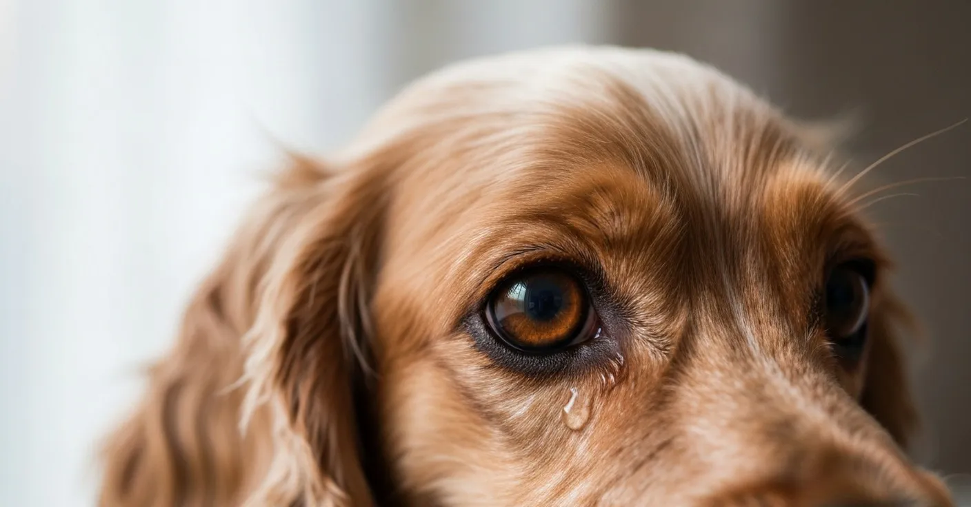
The first call I took on a Saturday morning a few years back was from a woman who’d woken up to find her Cocker Spaniel’s right eye glued shut with what she described as “yellow crust.” She was panicked, and she was right to be. By the time she got to the clinic an hour later, the eye was visibly swollen and the dog was holding it half-closed against the light. We started topical antibiotics that afternoon and treated a deep corneal ulcer over the following two weeks.
Two days later, I saw a Maltese in the same exam room whose owner had been worrying for six months about a faint clear glaze under both eyes. That dog needed nothing. The discharge was normal tear overflow on a coat color that made it look more dramatic than it was.
Same word — “discharge” — but two completely different clinical pictures. Knowing which is which doesn’t replace a vet visit, but it does help you decide how urgent that visit needs to be. The color, the texture, whether one eye or both are involved, and what your dog is doing alongside it all matter.
How tear film and eye discharge actually work
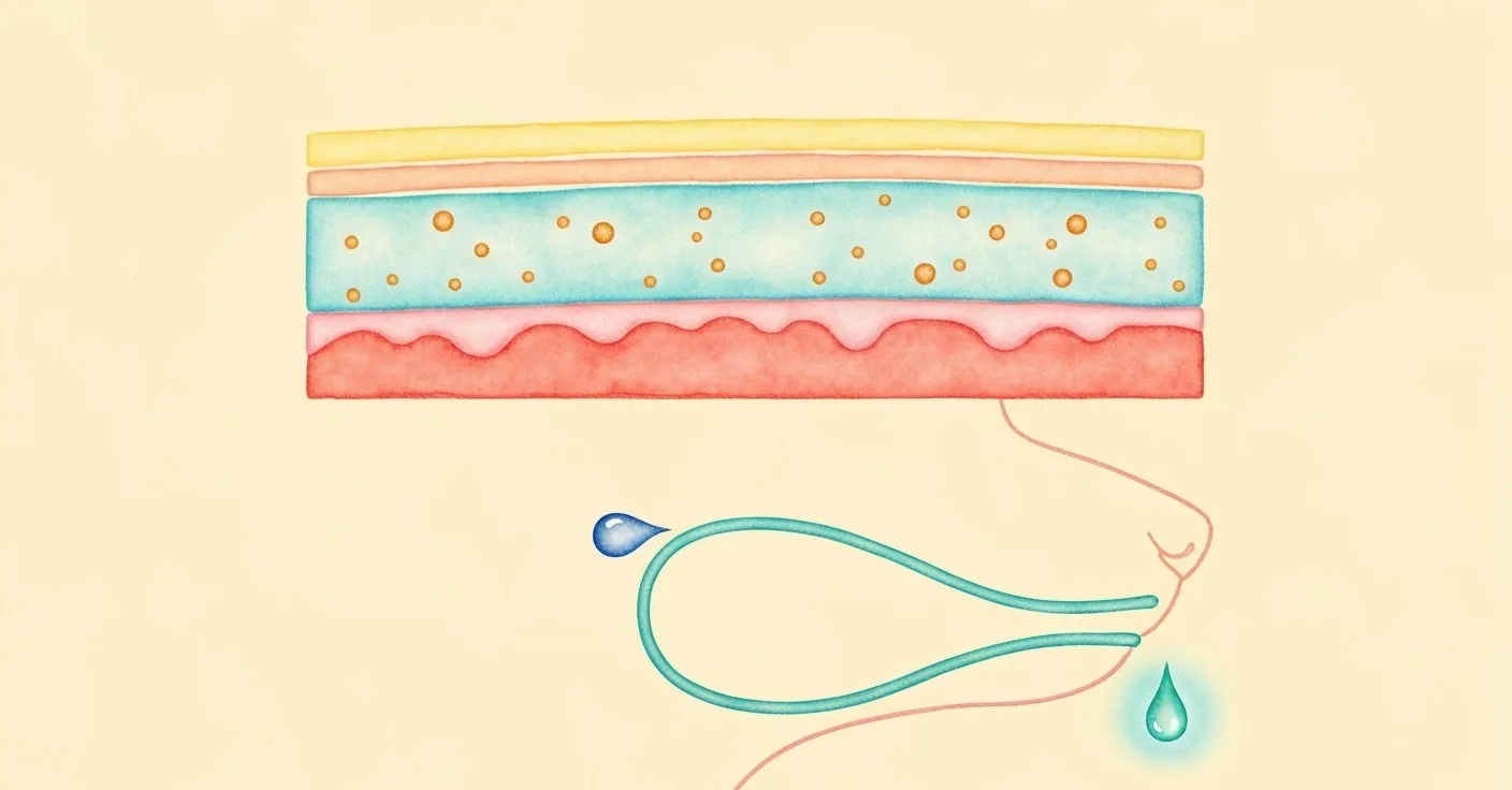
A dog’s eye is constantly producing tears, even when nothing looks wet. Tear film has three layers: a thin outer lipid layer that prevents evaporation, a thick middle aqueous layer that carries oxygen and nutrients to the cornea, and an inner mucin layer that helps the film cling to the corneal surface. Every blink redistributes this film across the eye, and the excess drains through tiny openings at the inner corner called the puncta, then down through the nasolacrimal duct into the nose (Maggs et al., 2013).
When this system is working, you don’t see discharge at all. A dog’s eyes look clean, the fur around them stays dry, and any tiny bit of debris that collects overnight wipes away easily in the morning.
Discharge appears when something disrupts that balance. Either tear production goes up, drainage goes down, the composition of the tears changes, or the eye starts producing things tears shouldn’t contain — mucus from irritation, pus from infection, blood from injury. The kind of discharge tells you which of those is happening.
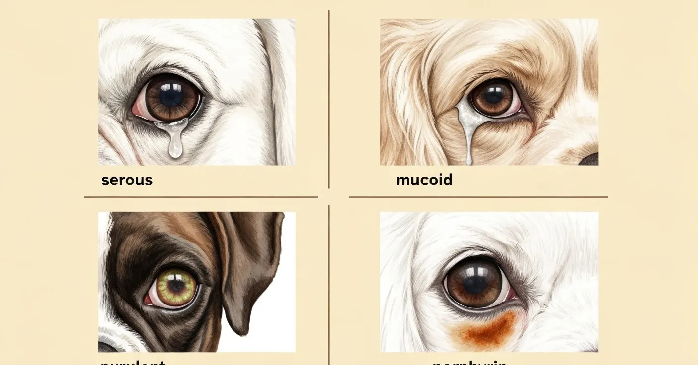
I find it helpful to think of discharge less as a single symptom and more as a category with four distinct subtypes, each pointing in a different direction.
Reading the color: what each type signals
Clear and watery
Clear watery discharge — what veterinarians call serous discharge — is mostly tear film. It can appear when tear production is higher than normal or when drainage is partially impaired and the tears overflow onto the fur instead of going down the duct.
Causes range from completely benign to mildly worrying. A dog who just ran through tall grass or rolled in dusty bedding will often show some clear discharge for an hour afterward; that’s the eye flushing irritants away as designed. Brachycephalic breeds like Pugs, Bulldogs, and Shih Tzus often have chronic mild overflow simply because their nasolacrimal ducts are kinked from their facial structure. The tears never make it down the drain.
What clear watery discharge is almost never a sign of, on its own, is infection. Bacterial and most viral infections produce thicker, colored material. If the discharge stays clear, the eye looks white and bright underneath, and your dog isn’t squinting, you’re usually looking at one of the benign causes (Petersen-Jones & Crispin, 2002).
The exception is allergies. Environmental allergies — pollen, mold, dust mites — can produce a watery discharge that’s persistent and bilateral, often paired with face-rubbing or paw-licking. That’s worth flagging to a vet, but it’s an outpatient conversation, not an emergency.
White or gray mucoid
A step up in viscosity. White or gray mucoid discharge — the kind that forms a stringy gel at the inner corner of the eye, or a crusty pale film overnight — usually means the eye is producing too much mucin relative to aqueous tears.
The most common cause in dogs is dry eye, properly called keratoconjunctivitis sicca or KCS. The aqueous-producing tear glands are damaged or under-active, so the eye compensates by ramping up mucin production. The result is a thick, sticky, pale discharge that owners often describe as “boogers” — and a cornea that’s chronically dehydrated underneath.
KCS is more common than most owners realize. It’s especially common in Cocker Spaniels, Cavalier King Charles Spaniels, Bulldogs, and West Highland White Terriers, and it tends to develop slowly enough that owners normalize the discharge before they realize the cornea is taking damage (Hartley et al., 2014). Untreated KCS can lead to corneal pigmentation, vascularization, and ulcers.
White mucoid discharge with squinting, redness, or a hazy-looking cornea is a same-week vet visit. A Schirmer tear test takes thirty seconds and tells the vet exactly how much tear production is happening; the treatment, typically a topical immunomodulator like cyclosporine, is straightforward once diagnosed.
Yellow or green purulent
This is the discharge that wakes me up at night when an owner describes it on the phone. Yellow or green discharge means pus — and pus means bacteria, white blood cells, and active inflammation.
The underlying causes vary. Bacterial conjunctivitis is the most common: an infection of the conjunctival membranes that line the inner eyelid and the sclera. Corneal ulcers — breaks in the surface of the cornea, often from trauma, foreign bodies, or untreated dry eye — almost always come with purulent discharge because bacteria colonize the damaged surface fast. Foreign bodies lodged behind the third eyelid will produce a thick discharge on the affected side within twelve to twenty-four hours.
The Cocker Spaniel I mentioned at the top had a corneal ulcer that had probably started as a minor scratch a day earlier. By the time we saw her, the eye was producing enough purulent material to crust shut overnight. With aggressive topical antibiotic therapy and an Elizabethan collar to stop self-trauma, she healed in about two weeks. But corneal ulcers can deepen quickly, and a deep ulcer can progress to perforation in a matter of days if left alone (Featherstone & Heinrich, 2013).
What makes purulent discharge a same-day-call situation isn’t the color alone — it’s the company it keeps. Squinting, holding the eye partially closed (called blepharospasm), redness of the visible eye surface, head-shyness, and any change in the appearance of the cornea itself all push the urgency higher. Green discharge plus squinting plus a dog who’s pawing at the eye is an emergency call, full stop.
Rust-brown porphyrin staining
The fourth type isn’t really discharge in the clinical sense — it’s the visible aftermath of normal tear overflow. Porphyrin is an iron-containing molecule that dogs excrete through their tear film. When tears sit on white fur long enough to oxidize in air and sunlight, the porphyrins turn that characteristic rust-mahogany color.
If you’re seeing brown staining without active wetness, swelling, redness, or squinting, you’re looking at a cosmetic concern, not a medical one. The Maltese I mentioned at the top is the classic example: bilateral, stable, chronic, and the dog is completely comfortable. The fur is stained but the eyes underneath are normal.
This doesn’t mean nothing’s going on. There’s almost always some low-grade tear overflow producing the porphyrin deposition, and reducing that overflow — through diet, water source changes, and supplementation — can lighten new fur growth over a period of weeks. For the underlying mechanism and the full management picture, see our detailed explainer on what porphyrin actually is and our broader guide to tear stain causes and solutions.
What porphyrin staining is not is purulent discharge. If the color is more orange-rust than yellow-green, the texture is dry rather than wet, and there’s no swelling or squinting, you’re almost certainly dealing with chronic overflow rather than infection.
What the texture and timing tells you
Color is the loudest signal but not the only one. A few other features are worth tracking before you call the vet.
Unilateral vs. bilateral. Discharge in both eyes simultaneously, of the same character, usually points to a systemic cause — allergies, breed-driven anatomical overflow, a viral upper respiratory infection. Discharge in one eye only is more concerning because it suggests something localized: a foreign body, a corneal injury, a duct obstruction, an eyelid abnormality. Unilateral purulent discharge in particular deserves a same-week vet visit even if your dog seems otherwise fine.
Onset speed. Discharge that appears suddenly over a few hours is more concerning than discharge that has been stable for months. Sudden onset signals acute injury or acute infection. Chronic stable discharge usually has a structural cause — drainage anatomy, breed conformation, low-grade dry eye — that has been there all along.
Time of day. Some dogs have noticeably more discharge in the morning because the eye dries out during sleep and produces mucus to compensate. Crusts that wipe away easily and don’t reappear during the day are usually benign. Discharge that re-accumulates throughout the day, in contrast, is active production and warrants attention.
Behavior changes. A dog whose eyes are uncomfortable will tell you. Squinting, holding the eye partially closed, pawing at the face, head-shyness, reluctance to come into bright light, or sudden tearfulness during meals are all signals that the eye itself hurts. Discharge plus behavior change is a much stronger trigger to call the vet than discharge alone.
I tell my clients to take a phone photo of the eye every morning for three days when they’re worried about discharge. The pattern over seventy-two hours — getting worse, staying stable, or fading — gives you and your vet far more information than a single snapshot.
What to do if you notice this
Watch and support at home if: the discharge is clear and watery, or it’s a faint rust-brown stain on a white-coated breed with no other symptoms. Your dog’s eye looks bright and white underneath, they’re not squinting, and they’re behaving normally. Gently wipe the fur with a damp cotton pad once or twice a day to remove debris and reduce oxidation on stained coats. Daily nutritional support that helps maintain healthy tear chemistry and gut function may contribute to lighter new fur growth over four to eight weeks. If the discharge stays mild and unchanged for a week, you’re likely in the chronic-overflow category. If it worsens at any point in those seven days, move to the next tier.
Call your vet within 24–48 hours if: the discharge is white, gray, or stringy and you notice your dog squinting, rubbing the face, or showing any sensitivity to light. The discharge is in one eye only and has appeared in the last 24–72 hours. There is mild redness on the white of the eye, or the eyelid appears slightly puffy. Your dog is otherwise eating, drinking, and behaving normally, but the eye looks different than it did a few days ago. Your vet may run a Schirmer tear test for dry eye, a fluorescein stain to check for corneal ulceration, or a culture if infection is suspected.
Seek veterinary care immediately if: the discharge is yellow, green, or thick and purulent. Your dog is holding the eye closed, squinting persistently, or pawing at it. The cornea itself looks cloudy, hazy, or has a visible spot or scratch. There is visible swelling of the eyelids or the area around the eye. The eye appears to bulge or sit differently than the other side. There is blood in the discharge or on the surface of the eye. These signs can indicate a corneal ulcer, glaucoma, foreign body, or deep infection, all of which can damage vision permanently within hours to days. Do not wait until morning.
Daily care and prevention
Most chronic mild discharge — the rust staining, the morning crusts on a brachycephalic breed, the slow trickle on a senior dog — responds to a consistent daily routine more than to any single intervention.
Gentle wiping with a soft damp cloth in the morning and evening removes accumulated material before it irritates the skin or oxidizes onto the fur. Plain warm water on a cotton pad is usually enough; medicated wipes are rarely necessary unless your vet has prescribed them. Avoid scrubbing the eyelid itself, and never use anything that contains alcohol or strong fragrance near a dog’s eye.
Keeping the hair around the eyes trimmed short helps in long-coated breeds. Long fur wicks tears outward onto the face, expanding the staining area and trapping moisture against the skin where it can cause secondary irritation. A monthly visit to a groomer who knows your breed, or careful at-home trimming with blunt-tipped scissors, makes a measurable difference.
Diet and water quality are worth thinking about for dogs with chronic porphyrin staining. Some owners see a real reduction in staining after switching to filtered or distilled drinking water, especially in regions with iron-rich tap water. Food sensitivities can also drive low-grade inflammation that increases tear overflow, though this is harder to confirm without a structured elimination trial.
For dogs with chronic mild overflow and visible porphyrin staining, a daily nutritional approach can help maintain tear chemistry and gut conditions associated with cleaner tear production. Petterm Tear Stain Powder combines lactoferrin, lutein, and probiotics — three ingredients that target porphyrin-related staining from different angles. For the science behind why these three are paired together, the porphyrin explainer linked earlier walks through each mechanism in detail.
None of this replaces a vet visit when discharge changes in character. It’s the daily baseline care for dogs who already have a clean bill of ocular health and are simply prone to mild overflow.
Frequently asked questions
Is some eye discharge normal in dogs? Yes. A small amount of clear or pale tan crust at the inner corner of the eye in the morning is normal in many dogs. It’s accumulated mucus and dust from overnight blinking patterns. If it wipes away easily and doesn’t recur through the day, it’s not a concern.
My dog suddenly has yellow discharge in one eye. What should I do? Call your vet today. Unilateral yellow or green discharge usually indicates an active bacterial infection or a corneal injury, both of which need prompt examination. Don’t try over-the-counter eye drops without a diagnosis; some human eye products contain ingredients that can worsen corneal damage in dogs.
Why does my dog have more eye gunk in the morning? Tear production drops during sleep and the eye produces more mucin to keep the cornea protected. The mucin accumulates overnight and gets wiped away with the first blinks of the day. As long as it doesn’t reappear during the day and there’s no squinting or redness, this is typically normal.
Can allergies cause eye discharge in dogs? Yes. Environmental allergies often produce a persistent, watery, bilateral discharge along with paw-licking, face-rubbing, or skin irritation elsewhere on the body. A vet can confirm with a physical exam and discuss whether antihistamines, dietary changes, or environmental control will help most.
My white dog has brown staining under both eyes. Is that infection? Almost certainly not. Brown staining on a white coat is usually oxidized porphyrin from chronic mild tear overflow. It’s a cosmetic concern, not a medical one, as long as the eye itself looks healthy and your dog is comfortable. The porphyrin explainer covers what’s actually happening biologically.
How do I tell the difference between conjunctivitis and a corneal ulcer? You usually can’t tell from home, which is why a same-day vet visit matters when purulent discharge appears. A fluorescein stain test takes thirty seconds in the clinic and lights up any break in the corneal surface — it’s the definitive way to distinguish surface infection from corneal injury. Treatment differs significantly between the two.
Should I use saline rinses on my dog’s eye? Plain sterile saline (the kind used for human contact lens care, without preservatives or additives) is generally safe for gently flushing debris from the eye surface. It’s most useful when you suspect dust or pollen got in. It’s not a treatment for active discharge, and it shouldn’t replace a vet visit when discharge is colored, persistent, or accompanied by squinting.
When to contact your veterinarian
Watch at home: Clear watery overflow that has been chronic and stable. Rust-brown porphyrin staining without active wetness, redness, or squinting. Small amounts of pale crust at the inner corner in the morning that wipe away easily and don’t return during the day. Daily gentle wiping, breed-appropriate grooming, and consistent nutritional support are appropriate for this picture.
Call your vet within 24–48 hours: New onset of white or gray mucoid discharge, especially if your dog is squinting or rubbing the face. Discharge that has appeared in one eye only and is changing in character. Mild swelling of the eyelids or visible redness of the conjunctival tissue. Chronic discharge that has suddenly increased in volume or changed in color. Persistent watery discharge paired with face-rubbing or seasonal allergy signs.
Seek same-day or emergency care: Yellow, green, or thick purulent discharge in one or both eyes. Discharge accompanied by squinting, blepharospasm, or pawing at the eye. Cloudiness, haziness, or visible damage to the corneal surface. Visible swelling of the eyelids or around the orbit. The eye appearing to bulge or sit differently than the other side. Any bleeding from the eye or visible foreign body. These signs can indicate corneal ulcer, glaucoma, deep infection, or trauma, all of which can threaten vision within hours.
References
- Maggs, D.J., Miller, P.E., & Ofri, R. (2013). Slatter’s Fundamentals of Veterinary Ophthalmology. 5th ed. Elsevier.
- Gelatt, K.N., Gilger, B.C., & Kern, T.J. (2013). Veterinary Ophthalmology. 5th ed. Wiley-Blackwell.
- Petersen-Jones, S. & Crispin, S. (2002). BSAVA Manual of Small Animal Ophthalmology. 2nd ed. BSAVA.
- Featherstone, H.J. & Heinrich, C.L. (2013). Ophthalmic examination and diagnostics. In Gelatt et al., Veterinary Ophthalmology, 5th ed., 533–613.
- Hartley, C., Williams, D.L., & Adams, V.J. (2014). Effect of age, gender, weight, and time of day on tear production in normal dogs. Veterinary Ophthalmology, 17(1), 1–7.
Researched and reviewed by the Petterm Editorial Team · Last reviewed May 2026
This article is for educational purposes and is not veterinary medical advice. Petterm products are not intended to diagnose, treat, cure, or prevent any disease. Results may vary. Always consult your veterinarian before introducing a new supplement, especially if your pet has an existing eye condition, takes medication, is pregnant, or is under 12 weeks old.